First aid advice for injuries to bones and muscles including fractures, dislocated joints, sprains and strains, spinal injuries, and recovery from a spinal injury.
An injury may be damage to your body. it’s a general term that refers to harm caused by accidents, falls, hits, weapons, and more. In the U.S., many people injure themselves per annum. These injuries range from minor to life-threatening. Injuries can happen at work or play, indoors or outdoors, driving a car, or walking across the road.
In our striated muscle injuries will represent a superb a part of all traumas in medicine, with an incidence from 10% to 55% of all sustained injuries. they need to be treated with the specified precaution since a failed treatment can postpone an athlete’s return to the world for weeks or even months and increase the danger of re-injury.
Topics in this section Injuries.
Introduction of Skeleton.
Introduction of Spine.
Introduction of Bones.
Introduction of Joints.
Introduction of Muscles.
Fractures.
Strains and Sprains.
Facial Injury.
Cheekbone and Nose Injury.
Lower Jaw Injury.
Collar Bone Injury.
Shoulder Injury.
Upper Arm Injury.
Elbow Injury.
Forearm and Wrist Injuries.
Hand and Finger Injuries.
Rib Injury.
Back Pain.
Hip and Thigh Injuries.
Fractured Pelvis.
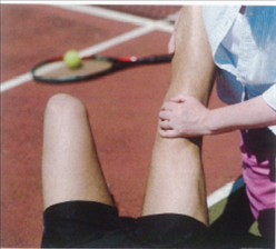
Knee Injury.
Lower Leg Injury.
Ankle Injury.
Foot and Toe Injury.
Cramp
What is the SKELETON?
The skeleton is the supporting framework around which the body is constructed. It is jointed in many places. and muscles attached to the bones enable us to move. Most of our movements are controlled at will and coordinated by impulses that travel from the via e nerves to every muscle and joint of the body, It is difficult for the first aider to distinguish between different bone, and muscle injuries, So this chapter begins with an overview of how bones, muscles, and Joints function and how damage occurs.
First aid treatments for most injuries from serious fractures to sprains, strains, and dislocations. are included here section, First aid for head and spinal injuries are covered in the chapter that deals with the nervous system because of the potential damage these injuries can have on the brain and spinal cord.
AIMS AND OBJECTIVES
- To assess the casualty’s condition quickly and calmly
- To steady and support the injured part of the body.
- To minimize shock
- Call 999/112 for emergency help if you suspect a serious Injury.
- To comfort and reassure the casualty
- To be aware of your own needs.
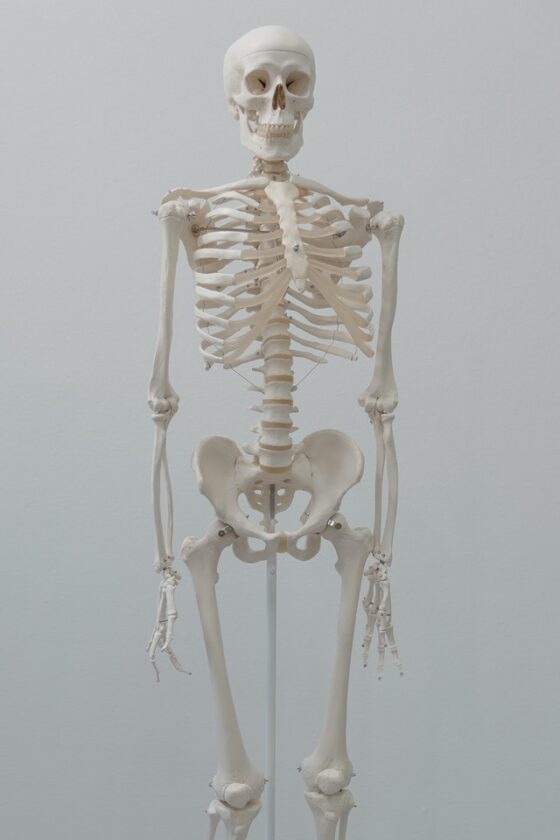
THE SKELETON
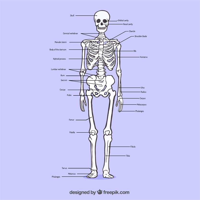
The body is built on a framework of bones called the skeleton. This structure supports the muscles, blood vessels, and nerves of the body. Many bones of the skeleton also protect important organs such as the brain and heart. At many points on the skeleton, bones articulate with each other by means of joints. These are supported by ligaments and moved by muscles that are attached to the bones by tendons.
The skeleton
There are 208 bones in the skeleton, providing a protective framework for the body. The skull. spine and ribcage protect vital body structures the pelvis supports the abdominal and the Bones and Joints of the arms and legs enable the body to move.
THE SPINE
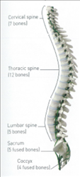
Also known as the backbone. the spine has a number of functions. It supports the head, makes the upper body flexible, helps to support the body’s weight, and protects the spinal cord. The spine is a column made up of 33 bones called vertebrae, which are connected by joints.
Between individual vertebrae are discs of fibrous tissue, called intervertebral discs. which helps to make the spine flexible and cushion it from jolls. Muscles and ligaments attached to the vertebrae help to stabilize the spine and control the movements of the back.
Spinal Column

The vertebrae form five groups: the cervical vertebrae support the head and neck: the thoracic vertebrae form an anchor for the ribs; the lumber vertebrae help to support the body’s weight and give stability: the sacrum supports the pelvis; the coccyx forms the end of the spine.

THE SKULL

This bony structure protects the brain and the top of the spinal cord. It also supports the eyes and other facial structures. The skull is made up of several bones, most of which are fused at joints called sutures. Within the bone are air spaces (sinuses), which lighten the skull. The bones covering the brain form a dome called the cranium. Several other bones from the eye sockets, nose. cheeks and jaw.
BONES, MUSCLES AND JOINTS
THE BONES
Bone is a living tissue containing calcium and phosphorus. minerals that make it hard. rigid and strong. From birth to early adulthood. bones grow by laying down calcium on the outside, They are also able to generate new tissue after injury. Age and certain diseases can weaken bones, making them brittle and susceptible to breaking or crumbling, either under stress or spontaneously. Inherited problems. or bone disorders such as rickets. cancer and infections. can cause bones to become distorted and weakened.
Damage to the bones during adolescence can shorten a bone or impair movement. In older people, a disorder called osteoporosis can cause the bones to lose density, making them brittle and prone to breaking.
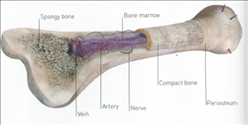
Part of a Bone
Each bones covered by a membrane (periosteum) which contains nerves and blood vessels. Under this membrane ‘s a layer of compact. dense bone, at the core is spongy bone in some bones, there is a cavity at the center containing soft tissue called bone marrow.
THE MUSCLES

Muscles cause various parts of the body to move. Skeletal (voluntary) muscles control movement and posture.
They are attached to bones by bands of strong, fibrous, tissue (tendons) and many operate in groups. As one group of muscles contracts, its paired group relaxes.
Involuntary muscles operate the internal organs. such as the heart. and work constantly, even while we are asleep. They are controlled by the autonomic nervous system.
Straightening the arm

The triceps muscle. at the back of the upper arm, shortens (contracts) to pull down the bones of the forearm. The basic muscle, at the front Of the arm, relaxes.
Bending the arm

The biceps muscle, at the front of the arm shortens (contracts) to pulling the bones of the upwards to bend the arm. At the same time. the triceps muscle relaxes and lengthens.
THE JOINTS
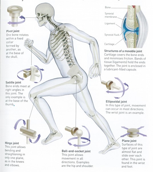
A joint is where one bone meets another. In a few joints (immovable joints) the bone edges fit together or are fused. Immovable joints are found in the skull and pelvis.
Most joints are movable and the bone ends are joined by fibrous tissue called ligaments, which form a capsule around the joint. The capsule lining (synovial membrane) produces fluid to lubricate the joint; the ends of the bones are also protected by smooth cartilage. Muscles that move the joint are attached to the bones by tendons. The degree and type of movement depends on the way the ends of the bones fit together. the strength of the ligaments and the arrangement of muscles.
FRACTURES
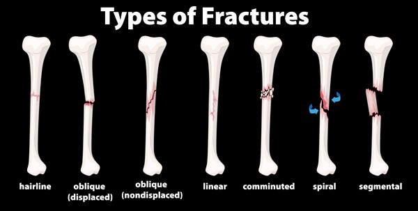
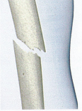
A break or crack in a bone is called a fracture. Considerable force is needed to break a bone. unless it is diseased or old. However. bones that are still growing are supple and may split. bend or crack like a twig. A bone may break at the point where a heavy blow is received. Fractures may also result from a twist or a wrench (indirect force).
RECOGNITION
There may be:
- Deformity, swelling, and bruising at the fracture site.
- Pain and or difficulty in moving the
- Shortening, bending, or twisting Of a limb.
- Coarse grating (crepitus) of the bone ends that can be heard or felt (by casualty)
- Do not try to seek this:
- Signs of a shock especially if the thigh bone or pelvis are fractured.
- Difficulty in moving a limb normally or at all If example. (inability to walk)
- A wound, possibly with the bone ends protruding [Treating an open fracture.
OPEN AND CLOSED FRACTURES
In an open fracture, one of the broken bone ends may pierce the skin surface. or there may be a wound at the fracture site.
An open fracture carries a high risk of becoming infected. In a closed fracture. the skin above the fracture is intact. However. bones may be displaced (unstable) causing internal bleeding and the casualty may develop shock.
Open fracture

Bone is exposed to the surface where it breaks the skin. The casualty may suffer bleeding and shock. Infection is rick.
Closed fracture

The skin is not broken. although the bone ends may damage nearby tissues and blood vessels, Internal bleeding a risk.
STABLE AND UNSTABLE FRACTURES
A stable fracture occurs when the broken bone ends do not hecause they are not completely broken or they are impacted. Such injuries are common at the wrist, shoulder, ankle and hip. Usually, these fractures can be gently handled without further damage.
In an unstable fracture, the broken bone ends can easily move. There is a risk that they may damage blood vessels, nerves, and organs around the injury Unstable injuries can occur if the bone is broken or the ligaments are torn (ruptured), They should be handled carefully to prevent further damage.
Stable fracture
Although the bone is fractured, the ends of the injury remain in place. The risk of bleeding or further damage is minimal.
Unstable fracture
In this type of fracture. the broken bone ends can easily be displaced by movement or muscle contraction.
TREATING A CLOSED FRACTURE
YOUR AIMS
- To prevent movement at the injury site.
- To arrange removal to hospital. With comfortable support during transport.
CAUTION
- Do not move the casualty until the injured part is secured and supported. unless she is in Immediate danger.
- Do not allow the casualty to eat or drink because an anesthetic may be needed.
- Advise the casualty to keep still. Support the joints above and below the injured area with your hands, or ask a helper to do this. until it is immobilized with a sling or bandages.

2. Place padding around the injury for extra support. Take or send the casualty to the hospital; an arm injury may be transported by car; call 999/112 for emergency help for a leg injury.
3. For firmer support and/or if removal to the hospital is likely to be delayed. secure the injured part to an unaffected part of the body. For upper limb fractures. immobilize the arm with a sting, For lower limb fractures. move the uninjured leg to the injured one and secure it with broad-fold bandages Always tie the knots on the uninjured side.
4. A Treat for shock if necessary. Do not raise an injured leg. Elevate an uninjured limb if shock is present. Monitor and record vital signs while waiting for help. Check the circulation beyond a sling or bandage every ten minutes. If the circulation is impaired, loosen the bandages.
SPECIAL CASE REALIGNING A LIMB
If a fractured limb bent and you need to immbbiliies it because removal to hospital will be delayed. it may be gently realigned to restore It to a suitable position. Pull gently in the line of the bone until the limb is straight. Pull only in a straight line and maintain in this position until the limb immobilised with bandages. Stop Immediately if traction increases the pain.
TREATING AN OPEN FRACTURE
YOUR AIMS
- To prevent blood loss. movement arid infection at the site of Injury
- To arrange removal to hospital with comfortable support during transport.
CAUTION
- Do not moue the casualty until the injured part is Secured and supported unless he is in immediate danger.
- Do not allow the casualty to eat or dank because an anesthetic may be needed.
- Do not press directly ort a protruding bone end.
- Cover the wound with a sterile dressing or a large, clean, non-fluffy pad. Apply pressure around the injury to control bleeding be careful not to press on a protruding bone.
- Carefully place a sterile wound dressing or more clean padding over and around the dressing.
- Secure the dressing and padding with a bandage. Bandage firmly, but not so tightly that it impairs the circulation beyond the bandage.

4. Immobilise the injured part for a closed fracture and arrange to transport the casualty to the hospital.
5. Treat the casualty for Shock if necessary, Do does not raise the injured leg. Monitor and record vital signs level of response. breathing and pulse while waiting for help to arrive. Check the circulation beyond the bandage every ten minutes. If the circulation is impaired, loosen the bandages.
SPECIAL CASE PROTRUDING BONE

It a bone end protruding, build-up pads at clean, soft. non-fluffy material around the bone. until you can bandage Over it without pressing on the injury.
DISLOCATED JOINT
This is a joint injury in which the bones are partially or completely pulled out of their normal position. Dislocation can be caused by a strong force wrenching the bone into an abnormal position or violent muscle contractions This very painful injury most often affects the shoulder. jaw or joints in the thumbs or fingers. Dislocations may be associated with torn ligaments or damage to the synovial membrane that lines the joint capsule.
Joint dislocation can have serious consequences. If vertebrae are dislocated, the spinal cord can be damaged. Dislocation of the shoulder or hip may damage the large nerves that supply limbs and result in paralysis. Dislocation of any joint may also fracture the bones involved. It is difficult to distinguish a dislocation from a closed fracture. If you are in any doubt. treat the injury as a fracture.
RECOGNITION
- “Sickening” severe pain.
- Inability to move the joint.
- Swelling and bruising around the affected joint. Shortening, bending, or deformity of the area.
YOUR AIMS
- To prevent movement at the injury site.
- To arrange removal to hospital, with comfortable support during transport.
CAUTION
- Do not try to replace a dislocated bone into its socket as this may cause further injury.
- Do not move the casualty until the Injured part secured and supported. unless she is In Immediate danger.
- For a hand or arm, injury removes bracelets. rings and watches In case of swelling.
- Do not allow the casualty to eat or drink because an anesthetic may be needed.
- If, for example, the casualty has a dislocated shoulder. advise the casualty to keep still. Help her to support the injured arm in the position she finds most comfortable.
2. Immobilise the injured arm with a Sling or use broad-fold bandages for a leg injury.
3. Place padding between the injured limb and the body. For extra support for an injured arm. place a broad-fold bandage around the sling and the body.
4. Arrange to take or send the casualty to hospital. Treat for shock if necessary. Monitor and record vital Signs while waiting for help to arrive.
5. Check the circulation beyond the bandages every ten minutes.
STRAINS AND SPRAINS
The softer structures around bones and joints – the ligaments. muscles and tendons can be injured in several ways. Injuries to these soft tissues are commonly called strains and sprains. They occur when the tissues are overstretched and partially or completely torn (ruptured) by violent or sudden movements. For this reason. strains and sprains are frequently associated with sporting activities.
Strains and sprains should be treated initially by the “RICE” procedure:
R – Rest the injured part:
I – Apply an Ice pack or a cold pad;
C – Provide Comfortable support;
E – Elevate the injured part.
This procedure may be sufficient to relieve the symptoms. but if you are in any doubt as to the severity of the injury. treat it as a fracture.
MUSCLE AND TENDON INJURY
Muscles and tendons may be strained, ruptured, or bruised, A strain occurs when the muscle is overstretched, it may be partially torn. often at the junction between the muscle and the tendon that joins it to a bone. In a rupture, a muscle or tendon is torn completely, this may occur in the main bulk of the muscle or in the tendon.
Deep bruising can be extensive in parts of the body where there is a large bulk of muscle. Injuries in these areas are usually accompanied by bleeding into the surrounding tissues. which can lead to pain. swelling and bruising
Muscle Tears

Vigorous movements may cause muscle fibers. such thé hamstring in the leg to tear. Muscle tears can cause severe pain and swelling.
Ruptured Tendon

The Achilles heel tendon attaches the calf muscle to the heel bone. It can snap after sudden exertion and may need surgery and immobilization.
LIGAMENT INJURY
One common form of ligament injury is a sprain. This is the tearing of a ligament at or near a joint. It is often due to a sudden or unexpected wrenching motion that pulls the bones in the joint too far apart and tears the surrounding tissues.
Sprained ankle
This is due to overstretching or tearing of a ligament – the fibrous cords that connect bones at a joint In this example, one of the ligaments in the ankle is partially torn.
RECOGNITION
- Pain and Tenderness.
- Difficulty in moving the injury part, especially if it’s a joint.
- Swelling and bruising In the area.
YOUR AIMS
- Reduce swelling and pain.
- To obtain medical help if necessary.
- Help the casualty to sit or lie down. Support the injured part in a comfortable position. preferably raised.

2. Cool the area by applying a cold compress. such as an ice pack or cold pad. to the injury. This helps to reduce swelling. bruising and pain.

3. Apply comfortable support to the injured part. Leave the cold compress in place or wrap a layer of soft padding, such as cotton wool, around the area. Secure it with a support bandage that extends to the next joint: for an ankle injury. the bandage should extend from the base of the toes to the knees.

4. Support the injured part in a raised position to help minimize bruising and swelling in the area. Check the Circulation beyond the bandages every ten minutes. If the circulation is impaired, loosen the bandages.

5. If the pain is severe, or the casualty is unable to use the injured part. arrange to take or send him to the hospital. Otherwise. advise the casualty to rest the injury and to seek medical advice if necessary.
FACIAL INJURY
Fractures of facial bones are usually due to hard impacts, Serious facial fractures may appear frightening. There may be a distortion of the eye sockets, general swelling and bruising, as well as bleeding from displaced tissues or from the nose and mouth. The main danger with any facial fracture is that blood, saliva or Swollen tissue may obstruct the airway and cause breathing difficulties.
When you are examining a casualty with a facial injury. assume that there is damage to the skull, brain, or neck. There is also a danger that you may misinterpret the symptoms of a facial fracture as a black eye.
RECOGNITION
- Pain around the affected area. If the jaw is affected, difficulty speaking. chewing or Swelling.
- Difficulty breathing.
- Swelling and distortion of the face
- Bruising and/or a black eye.
- Clear fluid or watery blood from the nose or ear.
YOUR AIMS
- keep the airway open.
- To pain and swelling.
- To arrange urgent removal to hospital.
CAUTION
- Never place a bandage around the lower part of the face or lower jaw case the casualty vomits or has difficulty breathing.
- Do allow the casualty to eat or drink because an anesthetic may be needed.
- If the casualty loses consciousness, open the airway and check breathing (The unconscious casualty)
- If an unconscious casualty is breathing. place him in the recovery position his Injured Side downwards so that blood or other body fluids can drain away Place soft padding under his Be aware of the risk of neck (spinal) injury.
- Call 999/112 for emergency help.
2. Help the casualty to sit down and make the airway is open and clear.
3. Ask the casualty to spit out any blood, displaced teeth, or dentures from his mouth. Keep any teeth to send to the hospital with him.
4. Gently place a cold compress against the casualty’s face to help reduce pain and minimize swelling. Treat for shock if necessary.

5. Monitor and vital signs level of response. breathing and pulse while waiting for help to arrive.
CHEEKBONE AND NOSE INJURY
Fractures of the cheekbone and nose are usually the result of direct blows to the face. Swollen facial tissues are likely to cause discomfort, and the air passages in the nose may become blocked. making breathing difficult. These injuries should always be examined in the hospital.
RECOGNITION
- Pain, swelling, and bruising.
- Obvious wound or bleeding from the nose or mouth.
YOUR AIMS
- To minimize pain and swelling.
- To arrange transport to the hospital.
CAUTION
- If there is clear fluid or watery blood leaking from the casualty’s nose. treat the casualty as for a head injury.
- Do not allow the casualty to eat or drink because an anesthetic may be needed.
- Gently place a cold compress. such as a cold pad or ice pack. against the injured area to help reduce pain and minimize swelling.
2. If the casualty has a nosebleed, try to pinch the nose to stop the bleeding Arrange to take or send the casualty to hospital.
LOWER JAW INJURY
Jaw fractures are usually the result of direct force, such as a heavy blow to the chin. In some situations. a blow to one side of the jaw reduces indirect force, which causes a fracture on the other side of the face. A fall to the point of the chin can fracture the jaw on both sides. The lower jaw may also be dislocated by é blow to the tace or is sometimes dislocated by yawning.
If the face is seriously injured. with the jaw fractured in more than one place, treat as for a facial injury (opposite).
RECOGNITION
- Difficulty speaking, swallowing and moving the jaw.
- Pain and nausea when moving the jaw.
- Displaced or loose teeth and drooling;
- Swelling and bruising inside and outside the mouth.
YOUR AIMS
- To protect the airway,
- To arrange transport to the hospital.
- If the casualty is not seriously injured. help him to sit with his head forward to allow fluids to drain from his mouth. Encourage the casualty to spit out loose teeth. and keep them to send to hospital with him.
2. Give the casualty a soft pad to hold firmly against his jaw in order to support it.
3. Arrange to take or send the casualty to the hospital. Keep his jaw supported throughout.
COLLAR BONE INJURY
The collar bones (clavicles) form “struts” between the shoulder blades and the top of the breastbone to help support the arms. It is rare for a collar bone to be broken by a direct blow. Usually. a fracture results from an indirect force transmitted from an impact at the shoulder or passing along the arm, for example. from a fall onto an outstretched arm. Collar bone fractures often occur in young people, as a result, of sports activities. The broken end of the collar bone may be displaced. causing swelling and bleeding in the surrounding tissues as well as distortion of the shoulder.
RECOGNITION
- Pain and tenderness. Increased by movement;
- Swelling and deformity Of the shoulder.
- Attempts by the casualty to relax muscles and relieve pain. she may support her arm at the elbow. and incline her head to her injured Side.
YOUR AIMS
- To Immobilise the injured shoulder and arm.
- To arrange transport to the hospital
CAUTION
- Do not allow the casualty to eat or drink because an anesthetic may be needed.
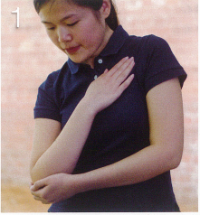
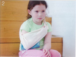
- Help the casualty to sit down. Lay the arm on the injured side diagonally across her chest with her fingertips towards her opposite shoulder. Ask her to support elbow the injured side with her other hand.
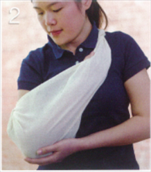
2. Support the arm on the affected side with an elevation sling.
3. Gently place Some Soft padding, such as a small towel or folded clothing, between the arm and the body to make the casualty more comfortable.
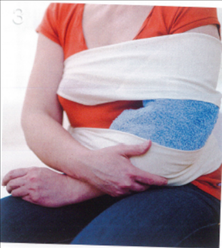
4. Secure the arm to the chest with a broad-fold bandage tied around the chest and over the sling. Once the arm is supported the casualty may be able to straighten her head.
5. Arrange to take or send the casualty to hospital in a sitting position.
SHOULDER INJURY
A fall on to the shoulder or an outstretched arm, or a wrenching force may pull the head of the upper arm bone (humerus) out of the joint socket – dislocation of the shoulder.
At the same time, ligaments around the shoulder joint may be torn. This injury can be very painful. Some people experience repeated dislocations and may need a strengthening operation on the affected shoulder.
A fall onto the point of the shoulder may damage the ligaments bracing the collar bone at the shoulder Other shoulder injuries include damage to the joint capsule and to the tendons around the shoulder: these injuries tend to be common in older people. To treat a shoulder sprain, follow the “RICE procedure” – Rest the affected part. cool the injury With Ice, provide Comfortable support with bandaging and Elevate the injury.
RECOGNITION
- Severe pain increased by movement: the pan may make the casualty reluctant to move:
- Åttempts by the casualty to relieve pain by supporting the arm and Inclining the head to the Injured Side.
- A flat, angular look to the shoulder.
YOUR AIMS
- To support and Immobilise the Injured limb.
- To arrange transport to the hospital.
CAUTION
- Do not attempt to replace a dislocated bone in Its socket.
- Do not allow the casualty to eat or drink because an anesthetic may be needed.
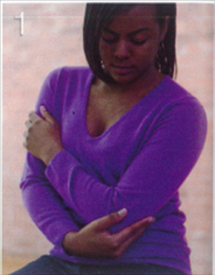
- Help the casualty to Sit down. Gently place the arm on the injured side across her body in the position that is most comfortable, with her hand towards the opposite shoulder. Ask the casualty to support her elbow on the injured side, or help her to do it.
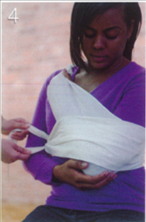
2. Support the arm on the injured side with an elevation sting make sure the knot is clear of the injury. Insert soft paddings, such as a folded towel or clothing. between the arm and the chest.
3. For extra support if necessary, Secure the limb to the chest by tying a broad-fold bandage around the chest and the Sling.
4. Arrange to take or send the casualty to hospital in a sitting position.
UPPER ARM INJURY
The most serious form of upper arm injury is a fracture of the long bone in the upper arm (humerus). The bone may be fractured across the center by a direct blow However, it is much more common, especially in elderly people, for the arm bone to break at the shoulder end. usually in a fall.
A fracture at the top of the bone is usually a stable injury as the broken bone ends stay in place. For this reason, it may not be immediately apparent that the bone is broken, although the arm is likely to be painful.
There is a possibility that the casualty will cope with the pain and leave the fracture untreated for some time.
RECOGNITION
- Pain, increased by movement;
- Tenderness and deformity over the site of a fracture.
- Rapid swelling.
- Bruising, which may develop more slowly.
YOUR AIMS
- To immobilise the arm.
- To arrange transport to the hospital.
CAUTION
- Do not allow the casualty to eat or drink because an anesthetic may be needed.
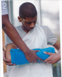
- Help the casualty to sit down. Remove all jewelry such as bracelets, rings, and Watches. Gently place the forearm horizontally across her body in the position that is most comfortable. Ask her to support her elbow if possible.

2. Slide a triangular bandage in position between the arm and the chest. ready to make an arm sling Place soft padding between the injured arm and the body. then support the arm and its padding in an arm sting.

3. For extra support, or if the journey to the hospital is prolonged. secure the arm by tying a broad fold bandage around the chest and over the sling; make sure that the broad-fold bandage is below the fracture site.

4. Arrange to take or send the casualty to the hospital.
ELBOW INJURY
Fractures or dislocations at the elbow usually result from a fall onto the hand. Children often fracture the upper arm bone lust above the elbow, This is an unstable fracture, and the bone ends may damage blood vessels. Circulation in the arm needs to be checked regularly. In any elbow injury. the elbow Will be stiff and difficult to straighten. Never try to force a casualty to bend it.
RECOGNITION
- pain, increased by movement.
- Tenderness over the site of fracture.
- Swelling. bruising and deformity.
- Fixed elbow.
YOUR AIMS
- To immobilise the arm without further injury to the joint
- To arrange transport to the hospital.
CAUTION
- lt the casualty feels faint, help her to lie down.
- Do not allow the casualty to eat or drink because an anesthetic may be needed.
- Do not try to move the injured arm.
- If the elbow can be bent, treat as for upper arm injury opposite. Remove all jewelry such as bracelets, rings, and watches.
2. If the casualty cannot bend her arm. help her to sit down. Place padding. such as a towel. around the elbow for comfort and support.
3. Secure the arm in the most comfortable position for the casualty using broad-fold bandages. Keep the bandages clear of the fracture site.
4. Arrange to take or send the casualty to the hospital.
5. Check the wrist pulse in the injured arm every ten minutes until medical help arrives. If you cannot feel a pulse, gently under the bandages and straighten the arm until the pulse returns. Support the arm in this position.
FOREARM AND WRIST INJURIES
The bones of the forearm (radius and ulna) can be fractured by an impact such as a heavy blow or a fall. As the bones have little fleshy covering. the broken ends may pierce the skin. produc•ng an open fracture.
A fall onto an outstretched hand can result in a fracture of the wrist. This is called a Colles fracture and commonly occurs in elderly people.
The wrist joint rarely dislocated but is often sprained, It can be difficult to distinguish between a sprain and a fracture, especially If the tiny scaphoid bone (at the base of the thumb) is injured. If you are in any doubt about the injury treats it as a fracture.
RECOGNITION
- Pan, increased by movement
- Swelling, bruising, and deformity.
- Possible bleeding with an open fracture.
YOUR AIMS
- To immobåilise the arm.
- To arrange transport to the hospital.
CAUTION
- Do not allow the casualty to eat or drink because an anesthetic may be needed.
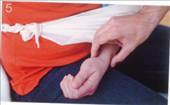
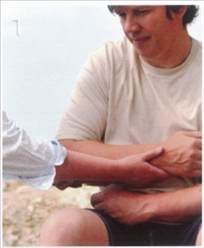
- Ask the casualty to sit down. Steady and support the injured forearm and place it across his body, ask the casualty to support it if he can. Expose and treat any wound.
2. Slide a triangular bandage in position between the arm and the chest. ready to make an arm sling. Surround the forearm in soft padding. such as a small towel.
3. Support the arm and the padding with an arm sling: make sure the knot is tied on the injured side.
4. For extra support or the journey to the hospital is likely to be prolonged. secure the arm to the body by tying a broad-fold bandage over the sling and body. Position the bandage as close to the elbow as you can. Arrange to take or send the casualty to the hospital.

HAND AND FINGER INJURIES
The bones and joints in the hand can suffer various types of injury. such as fractures, cuts and bruising. Minor fractures are usually caused by direct force A fracture of the knuckle often results from a punch, Multiple fractures. affecting many or all of the bones in the hand. are usually caused by crushing injuries, The fractures may be open, with severe bleeding and swelling. needing Immediate first aid treatment.
The joints in the fingers or thumb are sometimes dislocated or sprained as a result of a fall onto the hand (for example, while someone is skiing or ice skating). Always compare the suspected fractured hand with the uninjured hand because finger fractures result in deformities that may not be immediately obvious.
RECOGNITION
- The pain increased by the moment.
- Swelling. bruising and deformity.
- Possible bleeding with an open fracture.
YOUR AIMS
- Elevate the hand and immobilise it.
- Arrange transport to the hospital.
CAUTION
- Do not allow the casualty to eat or drink because an anesthetic may be needed.
- Help the casualty to sit down and ask her to raise and support the affected wrist and hand; help her if necessary. Treat any bleeding and loosely cover wound with a sterile stressing or large clean, un-fluffy pad.
2. Remove any rings before the hand begins to swell. and keep the hand raised to minimize swelling. Wrap the hand in soft. non-fluffy padding for extra protection.

3. Gently support the affected arm across the casualty’s body by placing it in an elevation sling.
4. For extra support or, if the journey to the hospital is likely to be prolonged. secure the arm by tying a broad-fold bandage around the chest and over the sling; keep it away from the injury. Arrange to take or send the casualty to the hospital.
RIB INJURY
One or more ribs can be fractured by direct force to the chest from a blow or a fall. or by a crush injury. If there is a wound over the fracture. or if a broken rib pierces a lung. the casualty’s breathing may be seriously impaired.
An injury to the chest can cause an area of fractured ribs to become detached from the rest of the chest wall. producing what is called a “flail-chest” injury. The detached area moves inwards when the casualty breathes in. and outwards as he breathes out.
This “paradoxical” breathing causes severe breathing difficulties. Fractures of the lower ribs may injure internal organs such as the liver and spleen, and may cause internal bleeding.
RECOGNITION
- Bruising. swelling or a wound at the fracture site.
- Pan at the site of injury.
- Pain on taking a deep breath.
- Shallow breathing
- A wound over the fracture.
- you may hear air being
- sucked” Into chest cavity.
- Paradoxical breathing.
- Signs of internal bleeding
YOUR AIMS
- To support the chest wall.
- To arrange transport to the hospital.
- Help the casualty to sit down and ask him to support the arm on the injured side; help him if necessary. For extra support place the arm on the injured side in a sling.
2. Arrange to take or send the casualty to hospital.
SPECIAL CASE A PENETRATING CHEST

If there a penetrating wound. help the casualty to sit down on the floor. leaning towards the injured Side. Cover and seal the Along three edges. Support the casualty with cushions and place the arm on the injured side in and call 999/112 for emergency help. Monitor and record Vital signs – level of response, breathing, and pulse while waiting for help.
BACK PAIN
Lower back pain is common and most adults may experience it at some point in their lives. It may be acute (sudden onset) or chronic (long term). It is usually caused by age-related degenerative changes or results from minor injury affecting muscles, ligaments, vertebrae. discs or nerves. It may be the result of heavy manual work, a fall or a turning or twisting movement. Serious conditions causing back pain are rare and bepnd the scope of first aid.
Most cases are simple backache, often in the lower back. in people aged 20-55 who are otherwise well. In a small number of casualties. the pain may extend down one leg. This is called sciatica and is caused by pressure on the nerve root la so-called “trapped nerve”
Spine injur•es in those under 20 or over 55, or that result from a more serious injury, require investigation and treatment and are dealt with in Nervous System Problems.
- Advise the casualty to stay active to mobiliser the injured area. Encourage him to return to normal activity as soon as possible.
- An adult casualty may take the recommended dose of paracetamol tablets. or his own painkillers.
3. Advise the casualty to seek medical advice if necessary.
HIP AND THIGH INJURIES
The most serious injury of the thigh bone (femur) is a fracture. It takes a considerable force, such as a car crash or a fall from a height. to fracture the shaft of the femur. This is a serious injury because the broken bone ends can pierce major blood vessels, causing severe blood loss, and shock may result.
Fracture of the neck of the femur is common in elderly people, particularly women. whose bones become less dense and more brittle with age (osteoporosis). This fracture is usually a stable injury in which the bone ends are impacted together. The casualty may be able to walk with a fractured neck of the femur for some time before the fracture is discovered. In the hip joint. the most serious. though much less common. type of injury is dislocation.
CAUTION
- Do not allow the casualty to eat or drink because an anesthetic may be needed.
- Do not raise the casualty’s legs. even if she shows signs Of shock. because you may cause further internal damage.
- Help the casualty to lie down. If possible, ask a helper to gently steady and support the injured limb at the knee.
- Gently straighten the casualty’s leg. If necessary, realign the limb, Support the limb at the ankle while you straighten it; pull gently in the line of the limb. Stop if this increases the pain.

3. Call 999/112 for emergency help. If the ambulance is expected to arrive quickly, keep the leg supported in the same position until it arrives.
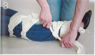
4. If the ambulance is not expected to arrive quickly, immobilise the leg by securing it to the uninjured one. Gently bring the sound leg alongside the injured one. Position a fold bandage at the ankles and feet Il), then a broad-fold one at the knees (2). Add additional bandages above (3) and below 141 the fracture site. Place soft padding between the legs to prevent the bony parts from rubbing. Secure the bandages on the uninjured side.

5. Take any steps possible to treat the casualty for shock: insulate her from the cold with blankets or clothing. Do raise her legs.
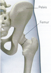
FRACTURED PELVIS
Injuries to the pelvis are usually caused by indirect force, such as a car crash, a fall from a height or by crushing. These incidents can force the head of the thigh bone (femur) through the hip socket in the pelvis.
A fracture of the pelvic bones may also be complicated by injury to tissues and organs inside the pelvis. such as the bladder and the urinary passages. The bleeding from large organs and blood vessels in the pelvis may be severe and lead to shock.
- Help the casualty to lie down on her back with het head flat/low to minimise shock. Keep her legs straight and flat or. if it is more comfortable, help her to bend her knees slightly and support them with padding, such as a cushion or folded clothing.
- Place padding between the bony points of the knees and the ankles. Immobilise the legs by bandaging them together with folded triangular bandages, secure the feet and ankles with a narrow fold bandage Ill, and the knees with a broad-fold bandage.

3. Call 999/112 for emergency help. Treat the casualty for shock Ipp. Do not raise the legs.
KNEE INJURY
The knee is the hinge joint between the thigh bone (femur) and the shin bone (tibia). It is capable of bending. straightening and, in the bent position. slight rotation.
The knee joint is supported by strong muscles and ligaments and is protected at the front by a disc of bone called the kneecap Ipatellal. Discs of cartilage protect the end surfaces of the major bones. Direct blows. violent twists or sprains can damage these structures. Possible knee injures include a fracture of the patella. sprains and damage to the cartilage.
A knee injury may make it impossible for the casualty to bend the joint, and you should ensure that the casualty does not try to walk on the injured leg. Bleeding or fluid in the knee joint may cause marked swelling around the knee,
- Help the casualty to Lie down. preferably on a blanket to insulate him from the floor or ground. Place soft padding. such as pillows. blankets or coats, under his Injured knee, to support it in the most comfortable position.
- Wrap soft padding around C the joint. Secure padding with a roller bandage that extends from the middle of the lower leg to mid-thigh.
3. call 999/112 for emergency help. The casualty needs to remain in the treatment position and so should be transported to the hospital by ambulance.
LOWER LEG INJURY
Injuries to the lower leg include fractures of the shin bone Itibial and the splint bone (fibula), as well as damage to the soft tissues (muscles ligaments and tendons)
Fractures of the tibia are usually due to a heavy blow for example, from the bumper of a moving vehiclel. As there is little flesh over the tibia, a fracture is more likely to produce a wound. The fibula can be broken by the twisting forces that sprain an ankle.
- Help the casualty to lie down, and gently steady and support the injured leg. If there is a wound, carefully expose it and treat the bleeding. Place a dressing over the wound to protect it.
2. Call 999/112 for emergency help. Support the injured leg with your hands; hold the joints above and below the fracture site to prevent any movement. Maintain support until the ambulance arrives.
3. If the ambulance is delayed, Support the injured leg by splinting it to the other leg. Bring the uninjured leg alongside the injured one and slide bandages under both legs. Position a narrow-fold bandage at the feet and ankles 11. then broad-fold bandages at the knees 12) and above and below the fracture site (3 and 4). Insert padding between the lower legs. Tie a figure-of-eight bandage around the feet and ankles, then secure the other bandages, knotting them on the uninjured side.

4. A If the casualty’s journey to hospital is likely to be long and rough, place soft padding on the outside of the injured leg. from the knee to the foot. Secure the legs with broad-fold bandages as described above.
ANKLE INJURY
If the ankle is broken, treat it as a fracture of the lower leg (opposite). A more common injury is a sprain usually caused by a twist to the ankle. This problem can be treated by the RICE procedure: Rest the affected part, cool the injury with Ice. provide Comfortable support with bandaging. and Elevate the injury.
- Rest and support the ankle in the most comfortable position for the casualty. preferably raised.
2. Apply a cold compress, such as an ice pack or a cold pad. to the site to reduce swelling and bruising.
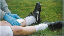
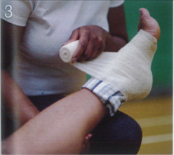
3. Apply comfortable support to the ankle. Leave the cold compress in place or wrap a layer of soft padding around the area. Bandage the ankle with a support bandage that extends from the base of the foot to the knee.
4. Raise and support the injured limb. Check the circulation beyond the bandaging. every ten minutes. If the circulation is impaired. loosen the bandage. Advise the casualty to rest the ankle and to seek medical advice if necessary.

FOOT AND TOE INJURIES
The bones and joints in the foot can suffer various types cf injury, such as fractures. cuts and bruising. Minor fractures are usually caused by direct force. Always compare the injured foot with the uninjured foot. especially toes. because
fractures can result in deformities that may not be immediate obvious. Multiple fractures. affecting many or all of the bones in the foot. are usually caused by crushing injuries.
These fractures may be open, with severe bleeding and swelling. needing immediate first aid treatment. Foot and toe injures must be treated in hospital.
- Help the casualty to lie down. and carefully steady and support the injured leg. If there a wound, carefully expose it and treat the bleeding. Place a dressing over the wound to protect it.
- Remove any foot jewelry before the area begins to swell.
- Apply a cold compress. such as an ice pack or a cold pad. This will also help to relieve swelling and reduce pain. A Place padding around the casualty’s foot and bandage firmly in place.

4. Arrange to take or send the casualty to hospital. If he is not being transported by ambulance. try to ensure that the injured foot remains elevated during travel.

5. Arrange to take or send the casualty to hospital. If he is not being transported by ambulance. try to ensure that the injured foot remains elevated during travel.
CRAMP
This condition is a sudden painful spasm in one or more muscles. Cramp commonly occurs during sleep. It can also develop after strenuous exercise. due to a build-up of chemical waste products in the muscles or to excessive loss of salts and fluids from the body through sweating or dehydration. Cramp can often be relieved by stretching and massaging the affected muscles.
CRAMP IN THE FOOT
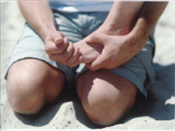
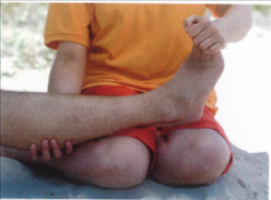
Help the casualty stand with his weight on the front of his foot: you can rest the foot on your knee to stretch the affected muscles. Once the spasm hag passed. massage the affected part of the foot with your fingers.
CRAMP IN THE CALF MUSCLES
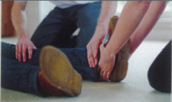
Help the casualty straighten his knee, and support his foot. Flex his foot upwards towards his shin to stretch the calf muscles, then massage the affected area on the back of the calf.
CRAMP IN THE FRONT OF THE THIGH

Help the casualty to lie down. Raise the leg and bend the knee to stretch the muscles. Massage he affected muscles once the spasm has passed.
CRAMP IN THE BACK OF THE THIGH
Help the casualty to lie down. Raise the leg and straighten the knee to stretch the muscles. Massage the area once the spasm has passed.